Displays images of the same side eye to compare them. Or select studies from previous dates to monitor progression.
The CX-1 is a Mydriatic Retinal Camera with full Non-Mydriatic functionality. Besides color photography, the CX-1 is equipped with high quality optical filters for FLUO, Red Free, Cobalt and standard even with FAF photography. The CX-1 can be changed into a NM camera by a simple push of a button. The Non-Mydriatic mode is essential for non dilatable patients such as glaucoma suspects. Children and photosensitive patients will also benefit from the non invasive IRED observation light. All photography modes can be performed in the MYD and NON MYD mode. This provides exceptional versatility and enables diagnosis, screening and monitoring of all major eye diseases
With the CX-1 following photography modes are available; Color, Red-free, Cobalt, Fluorescein Angiography and Fundus Autofluorescence (FAF) Alignment and observation with the CX-1 can either be done by the viewfinder or the large EOS LCD screen.

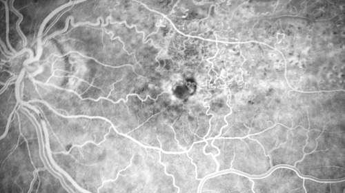
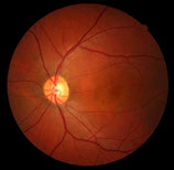
Clear, high resolution images are available in either mydriatic or non-mydriatic mode. The 2x mode magnifies the image by automatically cropping out the peripheral edges so the region of interest is larger in the frame. Canon Opacity suppression will largely suppresses the effects of cataracts and other ocular opacities.The image processing that is applied, reduces the gradation of overexposure. Low-intensity sections (macula) are clearly visible, while maintaining the gradation in tone of the high-intensity portion (the optic disc).
The image processing that is applied, reduces the gradation of overexposure. Low-intensity sections (macula) are clearly visible, while maintaining the gradation in tone of the high-intensity portion (the optic disc).
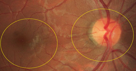
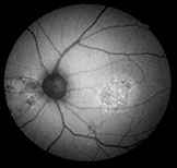
With the push of the Myd/Non-Myd button to select the non-mydriatic mode and the FAF mode, the operator is ready to begin taking images. The FAF mode helps monitor macular waste such as lipofuscin, which assists the eye care professional with detecting age-macular degeneration (AMD), glaucoma, and diabetes.
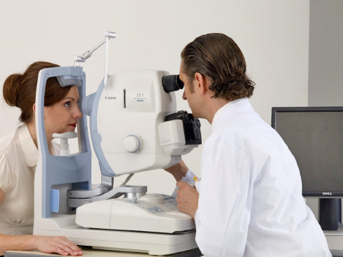
MYD Mode
Observation by viewfinder. Visible observation light.
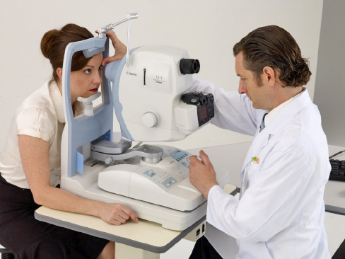
Non-MYD Mode
Observation by EOS screen. Invisible IR observation light.
FAF Imaging for the diagnosis of retinal disease is a relatively new diagnostic technique that provides more information on the health of the retinal pigment epithelium. FAF has proven to be very useful for the early detection of age related Macula Degeneration (AMD), one of the leading causes of visual impairment. Recent studies indicate that FAF Imaging can also aid in the diagnosis of a variety of other diseases and even in the detection of intraocular tumors.
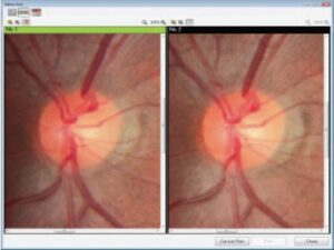
The LCD monitor displays guides which automatically determine the base length for acquiring successful stereo images. The two images can be stored as a pair making it easier to manage the files.

A single push of the Myd/Non-Myd button switches between the mydriatic and non-mydriatic imaging modes.
Whether the CX-1 is in mydriatic or non-mydriatic mode, the control panel has simple operation, workflow efficiency, and ergonomic controls.
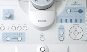
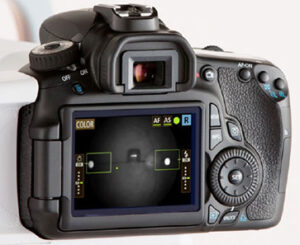
The EOS Retina camera provides optimal retinal imaging. And it is completely integrated with the functions of the retinal camera. A further advantage is that the digital camera body can be exchanged easily in case of upgrading to a newer model or service requirements.
Software designed to work with all Canon retinal cameras; for capturing, reviewing and reporting. When the camera is used stand-alone, it also functions as database with an archive function.
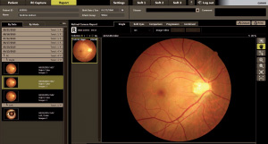
Extremely intuitive user interface. Clear graphic representation of functions and display of previous studies.
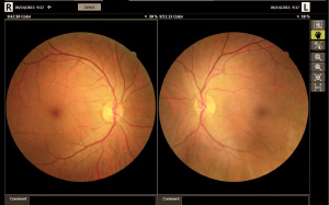
Displays images of the same side eye to compare them. Or select studies from previous dates to monitor progression.
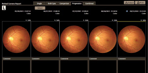
Observation progression: select up to 5 past examinations to see changes over time.
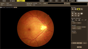
Report screen with extensive software tools: embossing a retinal image, changing gamma value, brightness and contrast, color balance balance, adding annotations and measuring C/D ratio.
EMBOSS can be useful to enhance the topography of the retina in the evaluation of macular degeneration, glaucoma, and retinopathy.
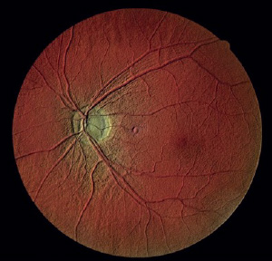
This function will make the blood vessels stand out.
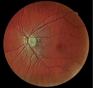
The optic disc stands out.
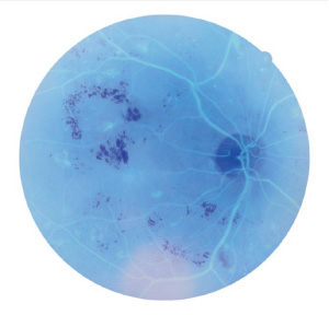
Inverts the color of an image to assist diagnosis.
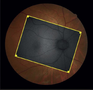
Overlay 2 images to see differences and changes in pathology. Selected images are aligned automatically. It can be useful for comparison to see changes, but also the colour image could be overlayed with e.g. a FAF image for additional information.
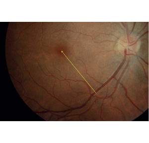
Add shapes, lines, arrows and comments to a captured image.
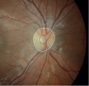
Measure the optic nerve papillary area. The C/D can be measured as line or area ratio.
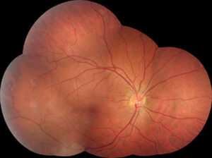
With the optional mosaic software up to 20 images can be combined, providing wide field imaging.
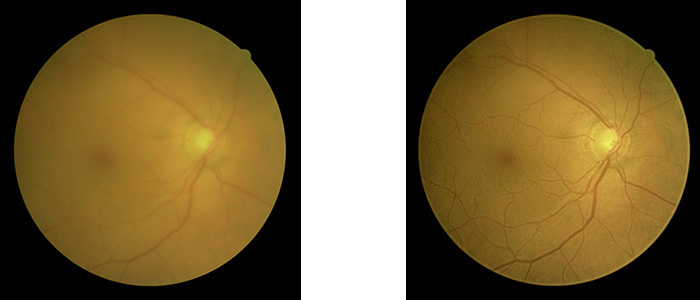
When obtaining retinal images, ocular opacity can cause several problems: the scattering of the light will make the edges of the blood vessels appear blurred, the difference in brightness of the retina will be reduced, making it very difficult to distinguish between structures. Furthermore a cataract eye will cause images to appear more yellow. Canon opacity suppression tool is a unique and sophisticated software tool, that based on the full information from the CMOS sensor of the digital camera, will largely suppress the effect of ocular opacities on color images.
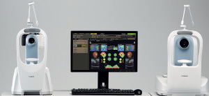
Our retinal cameras can be combined with a Canon OCT, using the same software and sharing the database. Output of both devices can be combined in one report to facilitate a comprehensive analysis.
Have questions? Send us a message about this product
Lenscan Medical Inc. provides a full scope of ophthalmic equipment, optical store equipment, dental loupes and surgical microscopes etc. to meet the needs of health care professionals. Lenscan Medical Inc. is committed to providing our customers highest possible level of service and support on our quality products.
© 2024 Copyright Lenscan Medical Inc.
Search Products